
What Is Fluoroscopy?
Fluoroscopy is an imaging method that uses continuous X-ray views to show movement inside the body on a monitor. A clinician can watch bones, joints, or injected contrast material in real time. The live images help guide certain procedures and help show how body structures move during an examination.
Quick Facts About Fluoroscopy
Category
Real-time X-ray imaging
Used for
Guided procedures and motion assessment
Common confusion
It is not magnetic resonance imaging
Also called
X-ray guidance, Fluoroscopic imaging
Often discussed with
Imaging Referrals
Key Takeaways About Fluoroscopy
- Live X-ray views distinguish fluoroscopy from still-image exams.
- Clinicians can track movement during imaging or a procedure.
- Fluoroscopy is not the same as an MRI scan.
How Fluoroscopy Works
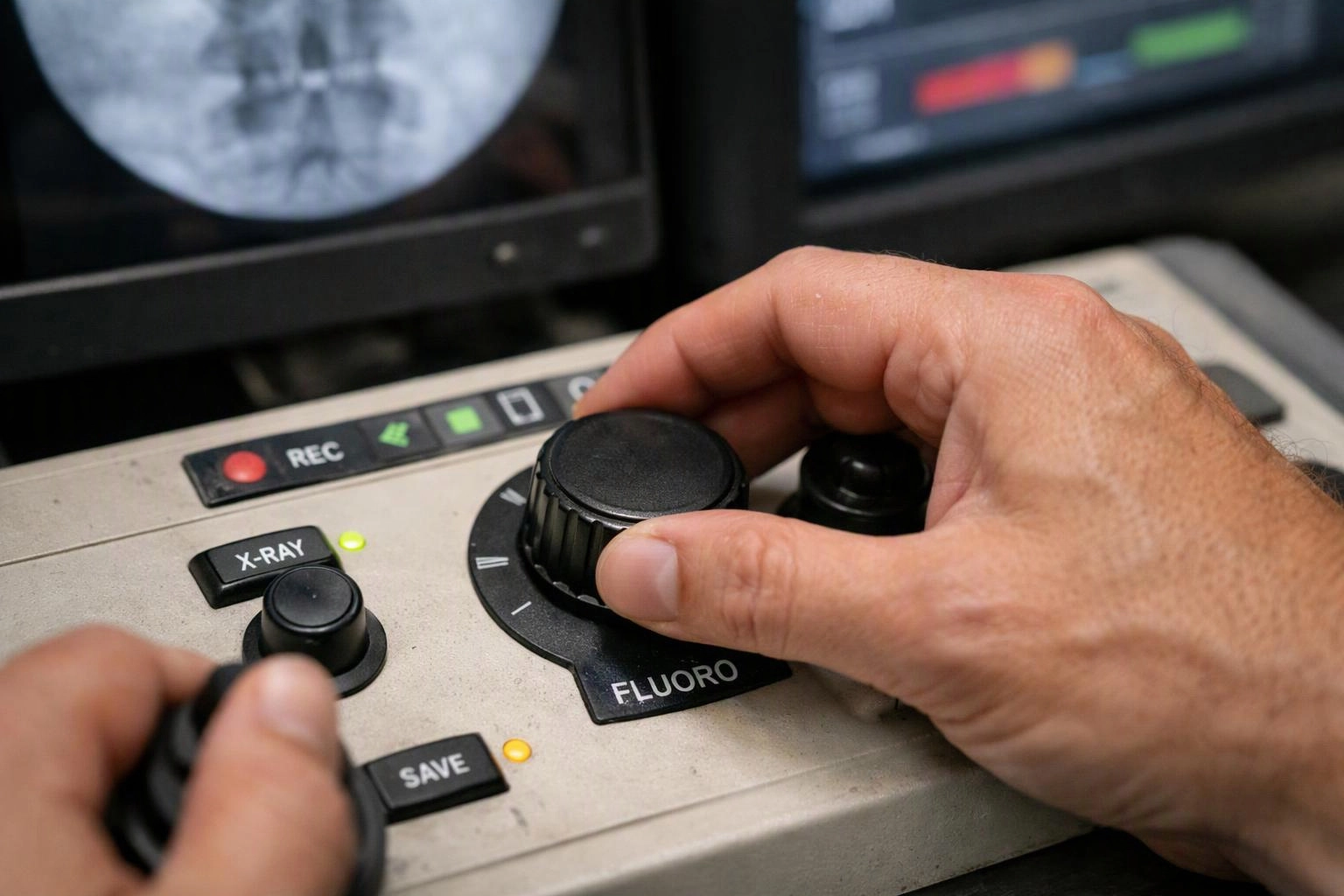
Fluoroscopy sends X-rays through the body and displays the images on a monitor. The moving view lets a clinician watch bones, joints, or contrast material as they change position. A still X-ray captures one view, while fluoroscopy shows motion during the exam.
Related glossary terms: Magnetic Resonance Imaging, Diagnostic Nerve Block, Epidural Space.
A fluoroscopy system includes an X-ray source and a detector. The detector turns the X-rays into images that appear on the screen. In pain care, the live view can help a clinician guide a needle toward a planned area. The image can also help confirm the target before medication or contrast material is placed.
Fluoroscopy may appear in an imaging referral when a referring clinician needs motion-based imaging or image guidance. The requested body area and clinical question help determine whether this method fits the referral.
From the Premier Injury Specialists team
When we coordinate Imaging Referrals, we review the clinical question and requested body area. We check whether fluoroscopy matches the type of imaging requested and pass along the relevant referral details. Our team also keeps the imaging method clear so the receiving facility understands the purpose of the exam.
Fluoroscopy in Practice: A Real-World Example
A person with persistent joint pain may receive an imaging referral for fluoroscopy when the clinician needs to observe joint movement. The live images can also guide a procedure aimed at the suspected pain source.
Related Services
Related Terms
Magnetic Resonance Imaging
Magnetic Resonance Imaging is a medical imaging method that uses a strong magnetic field and radio…
Diagnostic Nerve Block
A Diagnostic Nerve Block is an injection used to test whether a specific nerve or nerve…
Epidural Space
An epidural space is a narrow area inside the spinal canal that surrounds the outer covering…
Joint Aspiration
Joint Aspiration is a medical procedure that removes fluid from inside a swollen or painful joint…
Premier Injury Specialists
Have Questions About Fluoroscopy?
Contact Premier Injury Specialists for practical guidance on Fluoroscopy and related pain management physician work in McDonough.