
What Is Magnetic Resonance Imaging?
Magnetic Resonance Imaging is a medical imaging method that uses a strong magnetic field and radio waves to create detailed pictures inside the body. MRI images can show soft tissues, discs, nerves, joints, and the spinal cord. Doctors use these pictures to assess possible causes of pain, injury, or reduced movement.
Quick Facts About Magnetic Resonance Imaging
Category
Medical imaging method
Used for
Assessing soft tissue, spine, joint, and nerve concerns
Common confusion
MRI differs from X-ray and CT imaging
Also called
MRI
Often discussed with
Imaging Referrals, Spine Injury Evaluation
Key Takeaways About Magnetic Resonance Imaging
- MRI shows soft tissues and spinal structures in detailed images.
- A doctor reviews the images with your symptoms and examination.
- MRI is not the same as an X-ray or computed tomography scan.
How Magnetic Resonance Imaging Works
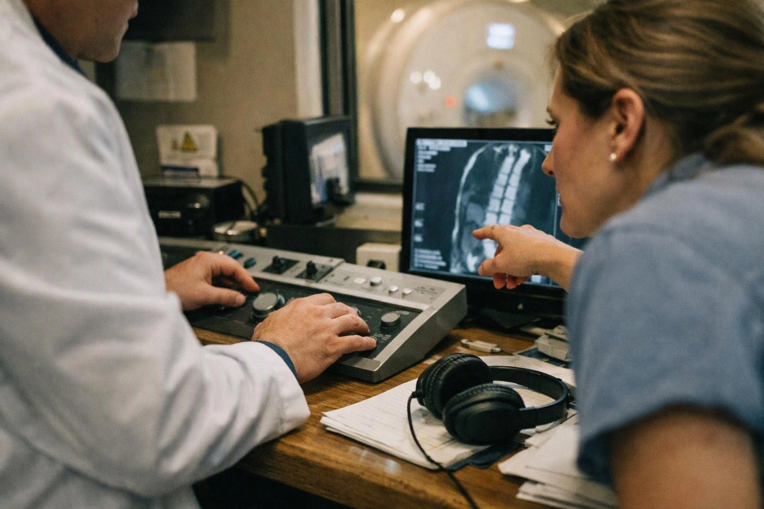
Magnetic Resonance Imaging uses a magnetic field and radio waves to produce images of internal body structures. The scanner collects signals from the body and turns them into cross-sectional pictures. A radiologist reviews the images and prepares a report for the ordering clinician.
Related glossary terms: Spinal Stenosis, Lumbar Radiculopathy, Annular Tear.
MRI can show soft tissues that may be difficult to assess on plain X-rays. These tissues include discs, ligaments, muscles, nerves, and the spinal cord. Radiology reports describe visible findings and help connect the images with symptoms and examination results.
How Imaging Referrals Use MRI Results
An imaging referral identifies the body area and the clinical concern being assessed. The receiving imaging center uses that information to plan the study and send the report to the appropriate medical team. MRI findings can support decisions about further evaluation or treatment.
From the Premier Injury Specialists team
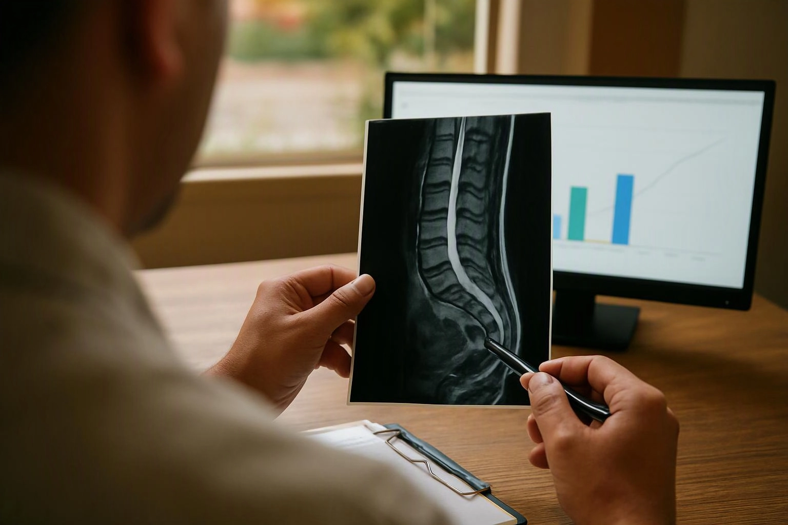
When we arrange an MRI referral, we match the requested body area with the patient’s reported symptoms and examination findings. We review the report with the rest of the medical record, since an image alone does not explain every pain complaint. We also track the report so it can inform the next clinical step.
Magnetic Resonance Imaging in Practice: A Real-World Example
A person with persistent lower back pain and leg symptoms may receive an MRI referral for the lumbar spine. The images can help the medical team assess discs, nerves, and other structures connected with those symptoms.
Related Services
Related Terms
Spinal Stenosis
Spinal Stenosis is a condition in which space inside the spinal canal or nerve openings becomes…
Lumbar Radiculopathy
Lumbar Radiculopathy is a nerve-related condition in the lower back that affects a nerve root near…
Annular Tear
An Annular Tear is a split or crack in the annulus, the tough outer ring of…
Disc Degeneration
Disc Degeneration is a gradual change in the spinal discs that reduces their water content and…
Premier Injury Specialists
Have Questions About Magnetic Resonance Imaging?
Contact Premier Injury Specialists for practical guidance on Magnetic Resonance Imaging and related pain management physician work in McDonough.